


The more complete and competent the diagnosis of prostatitis, the more effective the subsequent therapy will be. A formal approach by a doctor can result in a long and ineffective treatment for the patient. Your task is to identify the inflammation of the prostate and all the factors that cause it.
How doctors diagnose prostatitis
Prostatitis is diagnosed by a urologist or andrologist. After talking to the patient, the doctor prescribes the necessary tests: first a standard set (blood, urine, prostate secretion, rectal examination), then, if indicated, more detailed and high-tech methods are used: CT scan, MRImagnetic, ultrasound.
Taking anamnesis
During the initial consultation, the doctor will ask the following questions:
- Duration of sexual intercourse (if it got shorter, under what circumstances);
- Presence of discomfort in the groin during prolonged stay in a static position, as well as after drinking alcohol or hypothermia;
- Frequency and speed of urination (there is some difficulty, intermittent jet, you have to get up frequently to go to the bathroom at night);
- Quality of orgasm (still bright or blurry, pain in ejaculation).
The more details the patient remembers, the more complete the clinical picture made by the doctor.
Differential diagnosis
The symptoms of prostatitis are similar to those of a number of other diseases:
- Cystitis (cramps when urinating, pain in the lower abdominal region).
- Adenoma (difficulty urinating, feeling of heaviness in the groin).
- Prostate cancer (blood in the urine, problems with urination).
- Rectal pathologies: hemorrhoids, paraproctitis (inflammation), anal fissures, creptitis (ulcerative colitis).
Additional diagnostic methods and the rationale for their use are shown in Table 1.
Table 1. Differential diagnosis of prostatitis
| Disease | Risk group | Analysis |
|---|---|---|
| Hyperplasia | Men over 45 with no history of urethritis, catheterization, trauma to the bladder and urethra (circumstances that may explain the pain, blood in the urine) | Prostate ultrasound and digital examination |
| Prostatitis | Especially young men who have recently suffered from fever, hypothermia, in whose history there are no provocative factors (identical to hyperplasia) | ultrasound, complete blood count (CBC), digital prostate exam |
| Prostate cancer | Men over 45, with no history of provocative factors | Prostate ultrasound, PSA analysis, digital exam |
If necessary, other specialized doctors are involved in the diagnosis: proctologist, neurologist, vertebrologist. The last two experts identify the causes of pain associated with violation of the structure of the spine, violation of nerve endings.
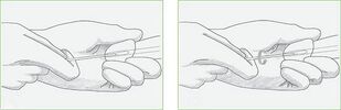
Rectal palpation
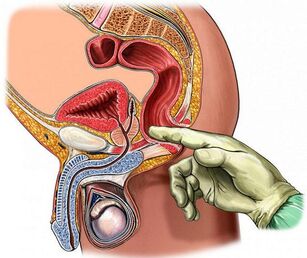
Digital rectal examination is the most accessible and informative method for checking the condition of the prostate. During the procedure, the doctor pays attention to the following parameters of its structure:
- Volume;
- Density;
- Surface roughness;
- Homogeneity (tissue homogeneity);
- Borders (outline clarity);
- Preservation of the isthmus (longitudinal suture between the lobes).
In prostatitis, the gland is enlarged due to edema (asymmetry is possible), its consistency is elastic, the longitudinal groove (suture) is not palpable and the patient may feel pain when touched.
For a clear picture for this type of diagnosisit is necessaryto prepare:
- Do not ejaculate the day before, do not drink alcohol, avoid heavy physical efforts, hypothermia and overheating.
- Don't ride a bike for a day, don't use rowing machines (don't hurt or massage your prostate in this way).
- Before consulting the doctor, perform an enema (a micro enema can be used) to clean the ampoule from the rectum.
You can feel the prostate at a depth of 3-5 cm from the anus. The doctor performs the procedure with sterile gloves, lubricating the finger with gel. The patient lies on his side with his knees bent or in the elbow position.
Laboratory methods
Laboratory methods to diagnose inflammation of the prostate involve the study of biomaterials for the presence of pathogens.
Blood
Based on the results of general and biochemical blood tests (take the capillary off a finger), prostatitis may be suspected at an early stage. The analysis is done on an empty stomach in the morning. You should avoid smoking an hour before the procedure.
Significant indicators:
- Leukocytes (blood cells, whose number increases with decreased immunity in the context of inflammatory reactions). Usually 4-9 × 10 ^ 9 units;
- ESR (erythrocyte sedimentation rate). The norm is around 5 units, an increase indicates inflammation or oncological process;
- Lymphocytes. Typically, their percentage of the total blood cell volume ranges from 18 to 40 units. Excess means infection.
Men over 40 are prescribed a PSA test- a tumor marker, the value of which exceeds what is meant by chronic prostatitis or prostate cancer.Norma- less than 4 ng / ml, after 50 years - 5, 53 ng / ml.
Urine
The urethra passes through the prostate (the prostatic part of the urethra), so when the gland becomes inflamed, the urine changes color and consistency. For the diagnosis of prostatitis, three types of analysis are performed:
- General - determination of physical and chemical parameters. Signs of inflammation of the prostate: urine is cloudy, whitish, alkaline, contains proteins, leukocytes, purulent filaments, sometimes foam or blood. In calculous prostatitis, phosphates are found.
- Cytological - exam for the presence of pathologically altered cells. The presence of erythrocytes and epithelium may indicate a tumor process.
- Bacteriological - identifies traces of the activity of pathogenic microorganisms. To do this, make a seeding tank in a nutrient medium. If there are bacteria and fungi, after a while they will start to multiply actively. Escherichia coli often causes prostatitis.
Before urinating, you should avoid eating salty and spicy foods, not consuming alcohol and dyes (beets, coffee). The analysis is done in the morning on an empty stomach.For prostatitis, the three-glass test method is used:the patient urinates alternately in each glass; the result is the first part, intermediate and final. This method allows you to identify the location of the inflammation: urethra, prostate, bladder. The four-pane method is more informative. The last urine sample is collected after massage of the prostate to obtain its secretion.
Secret of the prostate and sperm
The juice produced by the prostate is a valuable diagnostic material. Prepare for your fence in the same way as for a digital rectal exam. For the volume of the secret to be sufficient, you must abstain from sexual intercourse for three to five days.
Methods for examining prostate secretions:
- Microscopy;
- Backseeding;
- PCR (polymerase chain reaction).
PCR is the most accurate method. For the processing of biomaterials, special enzymes are used that multiply the number of pathogen DNA and RNA fragments. Research requires a special device - an amphiculator. Real-time PCR with greater precision. The result is ready in an hour.
Inflammation of the prostate is indicated by the presence in its juice of amyloid bodies, staphylococci, streptococci, Pseudomonas aeruginosa, epithelial cells (more than three units per field of view). The number of lipoid grains decreases and the number of leukocytes increases.
Thespermogram for prostatitis is an additional analysis. In the context of inflammation of the prostate, the sperm becomes yellowish or brown, its viscosity increases (liquefies for a long time) and the pathogenic microflora is present. In chronic prostatitis, gland epithelial cells, amyloid bodies and mucus are found.
Urethral swab
Urethral smear (scraping) is a less informative method for diagnosing prostatitis than secretion analysis.Used in cases where it is impossible to obtain the latter due to hemorrhoids, exacerbation of inflammation, presence of calcifications in the prostate body.
The procedure for removing the material is quick, but uncomfortable: the doctor dips a brush into the urethra, which captures part of the cells that cover it together with microorganisms. Then, the biomaterial is examined by PCR, which allows determining the presence of pathogens in any quantity. The cause of prostatitis can be genital infections: chlamydia, Trichomonas, mycoplasma.
Before doing the analysis for a day, you must refuse sexual intercourse in the morning, do only external hygiene procedures on the penis (do not spill anything in the urethra), do not urinate for two hours.
Instrumental methods
Instrumental diagnostic methods allow you to confirm and complement the results of laboratory tests.
Ultrasound and TRUS
The ultrasound examination of the prostate allows you to visualize its structure, contours, the nature of the tissue changes. In the case of prostatitis, transrectal ultrasound (TRUS) is considered the most informative: the doctor inserts the probe into the rectum. Prepare for the procedure in the same way as for palpation of the gland. An abdominal ultrasound (through the abdomen) is more comfortable for men, but the prostate is not completely visible due to the bladder.
With inflammation of the prostate, its structure is heterogeneous, the contours are blurred, there may be foci of fibrosis (enlarged connective tissue), scars. The prostate is dilated, the groove between its lobes is smoothed.
MRI, PET and CT
If the ultrasound gives reason to suspect the presence of a tumor process, the doctor prescribes CT (computed tomography) or magnetic resonance imaging (MRI) to clarify the condition. The latter type of research is more accurate, but also more expensive. The procedures are painless, in terms of informational content, they can replace a biopsy (tissue fragment clamping).
CT and MRI show in detail the structure of the prostate: calculi, cysts, tumors, inflammatory foci, structural abnormalities. For a clearer image, a contrast agent is preliminarily injected into the vein (not used in men with kidney failure). For the procedure, an appropriate type of CT scanner and rectal probe are used.
PET - positron emission CT. It allows you to analyze the condition of the prostate at the cellular and molecular level. It determines not only the presence and size of the tumor, but also the speed and quality of the metabolic processes that occur in it.
Regarding preparation:the rectum must be emptied. No food should be eaten for five hours before the procedure.
Characteristics of the diagnosis of certain types of prostatitis
Acute (infectious) bacterial prostatitis is diagnosed based on the patient's complaints, urinalysis, ultrasound, urethral smear. With active inflammation, the gland becomes sore, transrectal interventions are not allowed, in extreme cases, a careful examination of the finger.
Laboratory data for the diagnosis of acute prostatitis are not particularly informative. A urine culture may be advisable, but not necessary. With inflammation active, there is no time to wait for results. To alleviate symptoms, antibacterial therapy with broad-spectrum drugs is performed.
Chronic prostatitis practically does not manifest itself in any way, therefore, its detection requires a wide range of laboratory, physical and instrumental methods. Determination of the patient's immunological and neurological status may be necessary.
Palpation of the gland, urine and prostatic secretions are of utmost importance. The presence of more than 10 leukocytes in the field of view indicates inflammation. If the bacterial culture does not show the growth of the infectious microflora in the context of a high number of leukocytes, an analysis of genital infections will be necessary.
With the bacterial nature of inflammation, a large number of pathogens are found in urine and prostate juice. An undeniable microbiological sign of chronic inflammation: the number of microbes (CFU) is over 104 per ml. Some of them are numbered in dozens, so their presence in an amount of 10 to 102 per ml can indicate prostatitis.
In abacterial (non-infectious) inflammation, they are absent, but experts recommend, in these cases, a more in-depth analysis: prostate puncture, through which pathogens that live in closed prostate passages are extracted. At the same time, the bacterial culture is sterile, but the pathogen is still found in the end. Most of the time, it is one of the E. coli varieties.
Ultrasound does not always show chronic inflammation. In addition to the methods mentioned, the doctor can prescribe urofluxometry - measuring the flow rate of urine using special sensors.
Typical comorbidities
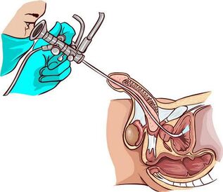
For long-term chronic prostatitis with signs of colliculitis (inflammation of the seminal tubercle near the prostate),urethroscopy is used - a visual examination of the canal using an endoscopic device. It helps to identify the narrowing of the urethra, violations of its structure, the state of the orifices of the excretory ducts of the prostate (mucus, pus, bulge) and the seminal tubercle.
Interpretation of results (definition of the stages of prostatitis by the state of the seminal tubercle):
- First: the seed tuber is red, edematous, bleeding. The same pattern is seen at the back of the urethra;
- Second: periodic increase and decrease in redness and swelling are characteristic;
- Third: scarring changes occur in the tissues of the tubercle and urethra, due to which the lumen of the ureter may narrow (stenosis).
Ureteroscopy irritates the receptors of the seminal tubercle, which compromises the microcirculation and motility of the prostate, so that the procedure is not performed unnecessarily.
Cystitis also accompanies chronic prostatitis. Inflammation of the bladder wall is detected using ultrasound and cystoscopy. During the research, pathological changes in the mucous membranes, mainly in the neck region, are determined. The state of the bladder in the context of chronic prostatitis (sclerosis of the prostate):
- Scar deformity of the bladder triangle.
- Dilated ureteral orifices.
- Narrowing of the neck.
Cystoscopy is prescribed in the final stage of the exam in the presence of lower abdominal pain and frequent urination.
The most difficult to diagnose is chronic abacterial prostatitis with pelvic pain of undetermined origin. In such patients, doctors, in the first place, conduct studies to exclude cystitis and neuropsychiatric pathologies.
How to diagnose prostatitis at home
A man may suspect acute prostatitis by the following signs:
- Severe pain in the lower abdomen and groin (between the testicles and the anus);
- Increased body temperature;
- Pain when urinating (such as cystitis);
- Premature and painful ejaculation.
The same symptoms appear during exacerbations of chronic prostatitis, caused by hypothermia or alcohol intake. The development of this form of pathology can be evidenced by the periodic appearance of blood in the urine, dull pain in the perineum (especially in a static position), difficulty in urinating, deterioration of the erection. These signs are the reason for contacting a urologist.
Conclusion
The longer the inflammatory process in the prostate lasts, the more difficult the treatment will be, so you should not delay the diagnosis. In government institutions, most follow-up procedures and treatments are free.